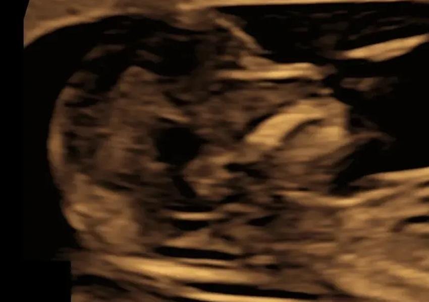
Third-month scan: NT-NB scan – Is it essential? Why and when?
Rekha (name changed) had come to me for a pregnancy scan for the second time. The first visit was for the early pregnancy scan. The tiny developing baby then was about the size of a kidney bean (7 weeks). The couple were overjoyed to see the baby’s heartbeat.
This time it was the NT scan visit. I briefly explained to her the purpose of the scan- screening for chromosomal abnormalities, screening for preeclampsia, looking at the baby's structural development, and so on. Rekha was still worried and wanted to know everything in detail. I probed- what was bothering her? She paused for a moment and told me about her elder sister’s son, who had been diagnosed with Down’s syndrome at birth. The child is 15 years now and is dependent on her parents entirely. Rekha’s eyes welled up while talking about the child. She loved the kid but felt sad that he could not be like kids of his age. Rekha wanted to know how Down’s syndrome could be detected while the baby was still in the womb.
All of us- treating doctors and expecting parents want the best outcome for a pregnancy. As the field of medicine is evolving, there is a shift from “diagnosing and treating” medical problems to “identifying risk factors and intervening” and thereby preventing a medical problem. The same rule is suitable for Fetal medicine as well. Several conditions can occur in the foetus and the mother that can be recognised early in pregnancy at about the third month, and that’s why the NT-NB scan is essential.
One of the conditions that affect the foetuses which can be detected prenatally (while in the mother’s womb) are chromosomal abnormalities. Chromosomes are microscopic thread-like structures that carry genetic information and are present in all body cells. Humans have 46 chromosomes, of which 23 come from the mother and the other 23 come from the father. Chromosomes determine how the different body structures develop and function. Errors in chromosomal makeup can lead to permanent changes in the system and functioning of the body and are not curable. The common chromosomal abnormalities are Down’s syndrome, Edwards syndrome, Patau’s syndrome. Down’s syndrome is the most common, and its incidence is thought to be 1 in 600 live births. Children with Down’s syndrome have an extra copy of chromosome 21, making 47 chromosomes in total. These babies can have structural problems with various body systems, have difficulties with neurodevelopment and can have learning disabilities.
The number of chromosomes in the foetus can be determined by taking a fluid sample around the baby and testing the cells present in the fluid for the total number of chromosomes. The procedure is done by inserting a thin needle into the uterus cavity and collecting the fluid- “amniocentesis”. However, this test cannot be done on all pregnant women as the above-said procedure is associated with a risk of a miscarriage of about 1 in 1000. This is the reason “screening” for chromosomal abnormalities have been developed.
Screening refers to application of a medical procedure or test to determine the likelihood of having a disease. The screening procedure itself does not diagnose the disease. It identifies “high risk” patients in whom the diagnostic test is performed to confirm/ refute the presence of disease.
Screening for chromosomal abnormalities -especially Down’s syndrome is performed between 11weeks to 13 weeks and 6 days of pregnancy. The test involves a scan called the NT-NB scan, and a blood test called double marker test, performed on the mother. The scan is performed to look for markers of chromosomal abnormalities. These are- NT- Nuchal translucency which is the fluid thickness at the back of the neck of the baby, NB- nasal bone, a bone found at the root of the nose. Other markers are blood flow across the tricuspid valve of the heart and flow across a blood vessel called ductus venousus. Most babies with Down’s syndrome have an increased Nuchal translucency or a non- ossified nasal bone with or without abnormal flow across the tricuspid valve and ductus venous. From the scan, based on the markers, a risk estimate is given which indicates the likelihood of baby having Down’s syndrome. However, some normal foetuses can also have these features and few babies with Down’s syndrome may not have any of these markers and may appear normal on scan. To increase the detection of babies with Down’s syndrome, the double marker test is performed on the mothers blood in addition to scan to look for 2 chemicals. One is a hormone called the Beta HCG (Human chorionic Gonadotropin) and the other is a protein called the (PAPPA Pregnancy Associated Plasma Protein A). The levels of these hormones are altered when baby is affected with Down’s syndrome. The risk assessment from the scan is combined with the blood test report to obtain a “combined risk” which gives an estimate of likelihood of baby having Down’s syndrome.
The final risk is categorised into high risk or low risk. When the risk is more than 1 in 100, eg., 1 in 60, it is considered high risk and when it is less than 1in 100, eg., 1 in 3000, it’s considered low risk. For women with low risk the chance of baby having Down’s syndrome is very low and further confirmatory test are not offered. For women with high risk, the baby's chance of having Down’s syndrome is high. Further, confirmatory diagnostic tests are offered which is amniocentesis- also called the needle test.
Detection rate (%) of Down’s syndrome based on the method of screening
Maternal age- 30%
Maternal age + Double marker: 60-70%
Maternal age + NT: 75-80%
Maternal age + NT+ Double marker: 85-95%
Maternal age + NT+ NB: 90%
Maternal age+ NT+ NB+ Double marker: 95%
REFERENCE: Nicolaides KH. Screening for fetal aneuploidies at 11 to 13 weeks. Prenatal diagnosis. 2011 Jan;31(1):7-15.
NT- Nuchal translucency
NB- Nasal bone

